Background

The accompanying primer, Thrombolysis for Stroke and role of CT perfusion Imaging, describes the difficulties and potential shortcomings of thrombolysis for acute stroke and the way that CT perfusion may improve patient selection for thrombolysis. This paper, by Hopyat et al. (Radiology 2010) describes a related problem: the risks of thrombolysis, mainly constituting secondary haemorrhage, are greater when reperfusing a large area of infarcted brain. In the second European Cooperative Acute Stroke Study (ECASS II), failure to recognise involvement of more than one-third of the middle cerebral artery territory resulted in a high risk of haemorrhage when such patients received thrombolysis. CT perfusion may allow better identification of this situation and avoidance of thrombolysis. In addition, CT perfusion may aid in identifying the baseline stroke size for prognostication and research purposes, in positive confirmation of ischaemia during a TIA and, as discussed in the primer, in identification of stroke mimics. The study uses an incremental protocol with up to date CT perfusion technology to assess its use in positive identification of stroke.
Study Design
The study took 191 consecutive patients with presumed stroke/ unresolved TIA who were admitted within 3 hours of symptom onset. Unenhanced CT, CT angiogram and CT perfusion were assessed in that order by non-expert reviewers. A final diagnosis of stroke was established about a month later by an experienced clinician with the aid of a subsequently-performed MRI with diffusion weighted imaging (DWI).
Results
According to the final diagnosis made retrospectively, 64% of the patients had stroke, 18% had TIA and 17% were stroke mimics.
The sensitivity, averaged over all patients and within and across image reviewers, of correct identification of stroke by unenhanced CT was 52.5%, by unenhanced CT and CT angiography was 58.3% and by unenhanced CT, CT angiography and CT perfusion all together was 70.7%; using all three was significantly better than using one or two modalities (p=0.0003 and p=0.013 respectively).
This was not at the cost of reduced specificity (i.e. false positive errors), which was around 85% for all three conditions. Rather than give an all or none answer, the reviewers scored their confidence levels for diagnosis of stroke, and this allowed calculation of receiver operating characteristic (ROC) curves for unenhanced CT alone, unenhanced CT and CT angiography and all three together.
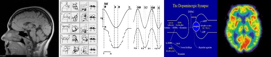
Receiver operating characteristics plotting sensitivity against false positive rate (i.e. 100-specificity) determined by reviewers scoring their confidence level in diagnosing stroke in various different patients. Unenhanced CT alone is the blue trace, unenhanced CT plus CT angiography is in brown, and unenhanced CT plus CT angiography plus CT perfusion is in orange.
For example a reviewer at a very conservative level, requiring a high confidence level for positive diagnosis, may rarely identify stroke; he will have high specificity when he does label a case as having a stroke, but very low sensitivity. A good diagnostic tool will be one where there is a larger area under the curve of sensitivity versus 100% minus specificity. In other words, accepting just slightly less than 100% specificity makes the sensitivity rise very dramatically. It is seen that using all three modalities together improves sensitivity over unenhanced CT alone at all levels of specificity, but at very high levels of specificity, CT perfusion does not improve performance over CT angiography.
Inter-observer agreement (Cohen kappa) was only between 0.28 and 0.44 for unenhanced CT alone, and between 0.68 and 0.78 for all three modalities together. Intra-observer agreement was similarly better using all three modalities together.
Strengths and Weaknesses of Study
The authors attempted a “real-life” situation analysis using an incremental protocol, a realistically early time of imaging, inexperienced reviewers and a range of stroke severities that included mild stroke and TIAs. They demonstrate clear superiority in these circumstances. The circumstances also explain why absolute performance may have been lower than in other studies.
The authors cite the advantages of CT perfusion, namely being done just after standard CT and taking just 1-2 minutes extra to perform and about 5 minutes extra to process. In their hands the radiation dose was only that of another unenhanced CT head. The disadvantages they cited are the confounding effects of chronic internal carotid artery occlusion and chronic ischaemic changes making it hard to determine what is new and what is old.
They consider the addition of CT perfusion well adapted to triage of stroke patients but are cautious about the benefits of identifying penumbra because of the absence of actual evidence that reperfusing penumbra improves outcome.
However, the “real-life” analysis situation might not be without shortcomings in interpretation because real life may be different in different units. Certainly in many stroke units there will be individuals on hand to assess imaging in real-time who may have several years’ experience rather than the one year’s experience of the study’s reviewers. Their lack of skill may have overestimated the extra sensitivity of CT perfusion.
While it is also laudable that they have not selected for their study only patients whose stroke was clinically obvious on admission, it does seem strange that there were so many TIAs when most TIAs do not usually last more than an hour. The mean delay was 117 minutes +/- 59 minutes, so generally there was a 1 to 3 hour window. Some clinicians might delay imaging a little if the patient attended within an hour with as yet not improving symptoms. It would not be a fault of imaging, as such, if a TIA was identified as stroke simply because the scan was performed early enough to detect the ischaemia. A more experienced radiologist might better distinguish ischaemia from established infarction on CT perfusion by the lack of reduction of cerebral blood volume, but this was not specifically examined in this study.
As a tool for ruling out stroke mimics, CT perfusion is clearly and unsurprisingly better than unenhanced CT (which was never intended for this purpose). But with sensitivities around 75% and specificities around 85%, it can hardly be considered a gold standard. Should we thrombolyse on that basis, given the 6% risk of causing harm from intracranial haemorrhage (though the harm engendered on thrombolysing the normal brain of some stroke mimics is likely to be low compared to thrombolysing an extensive established infarct)?
The performance of CT perfusion in positive diagnosis might in fact come up short compared with that of an experienced clinician examining the patient shortly after initial triage, and then one wonders whether that clinician ought not then to rely on his clinical judgement alone or upon an early MRI with DWI, accepting that this might not be universally feasible.
The introduction in the paper started by describing early major middle cerebral artery infarction as a relative contraindication for thrombolysis and how CT perfusion may help identify this. I cannot help but wish this is what the study actually investigated rather than detection of stroke mimics, but it does at least provide a good guide as to how to go about conducting such an investigation rigorously.

Well, that’s a lot of infor on CT perfusion! Guess there’s still no substitute for clinical skills. Thanks for the report.