 Background
It has long been known that there exist variants of Parkinson’s disease (PD), loosely and perhaps inaccurately described as PD plus syndromes, that may carry features of Parkinsonism but which also have other clinical features. Such conditions have distinct pathology at autopsy.
However, it has also long been known that clinicopathological correlations of these conditions are not perfect; in other words, a patient in life may have clinical features indicating one PD plus syndrome but may be found subsequently to bear the pathology of another.
The subject of this journal club, When Dementia with Lewy Bodies, Parkinson’s Disease and Progressive Supranuclear Palsy Masquerade as Multiple Systems Atrophy by Koga et al. (2015) in Neurology, is a retrospective review of Mayo Clinic brain bank cases labelled as having Multiple Systems Atrophy (MSA) in life.
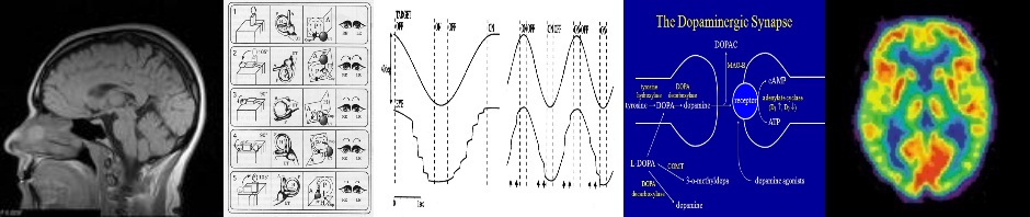
MSA is a neurodegenerative condition that may have one or both of parkinsonism and ataxia features, and may also have autonomic features, pyramidal features and even features of anterior horn cell disease. According to the Second Consensus Statement (2008), the criteria for probable MSA are:
A sporadic neurodegenerative condition of onset >30 with:
- Urinary incontinence (plus erectile dysfunction in males) or measuredorthostatic hypotension within 3 min of 30mmHg systolic or 15mmHg diastolic and at least one of:
- Poorly levodopa responsive Parkinsonism (bradykinesia with rigidity, tremor or postural instability)
- Cerebellar syndrome
However some patients will have pathologically proven MSA without satisfying these criteria, while in others the clinical picture will be confused by coexisting conditions in this age group, such as Alzheimer’s disease (AD) or cerebrovascular disease.
Study Design
The study reviewed the autopsy results of 134 cases that had consecutively been submitted to the brain bank with a clinical label of MSA. Patients came from 37 US states. The pathological assessments were done using a standard protocol. In 125 patients there were useful clinical records, and in some cases further information was gained by questionnaires sent to living relatives.
Study Findings
A pathological diagnosis of MSA was confirmed in 62% of cases. Of the remaining 38% of cases, 37% had Dementia with Lewy body (DLB) pathology, 29% had PSP, and 15% had PD. Two of the 134 total had Corticobasa Degeneration (CBD), two had cerebrovascular disease and five were “miscellaneous”.
On retrospective assessment of clinical features according to the above criteria, only 49 patients had probable MSA and 35 possible. (But incomplete records do not mean that patients did not have particular clinical features). Once this had been done, 71% of probable MSA patients had pathological MSA, and 60% possible MSA patients had MSA pathology.
The paper describes pathological changes in some detail. In the same way that there are, according to Braak, “stages” or at least grades of neurofibrillary tangle involvement in Lewy body disease, there have been described five phases of A beta amyloid deposition in Alzheimer-type disease. These range from phase 1 where deposition is exclusively in neocortex, to phase 5 where there is widespread involvement even in the cerebellum.
In pathological MSA, 8% also had Lewy body pathology. Overall the median Braak stage was I (not 0). A quarter of the MSA brains had phase 1 or worse A beta phase of Alzheimer’s.
With pathological diagnosis as the reference point, the features that were more common in MSA than in DLB were urinary continence, ataxia, nystagmus and pyramidal signs. Cognitive impairment and visual hallucinations were more common in the latter.
Comparing MSA vs PD, incontinence was more frequent and visual hallucinations less frequent.
Comparing MSA vs PSP, urinary incontinence, constipation, orthostatic hypotension and REM sleep behaviour disorder were more frequent, and vertical gaze palsy less frequent.
Levodopa responsiveness and mini-mental state score actually did not distinguish these diagnoses.
The main errors related to assuming that orthostatic hypotension automatically resulted in an MSA diagnosis instead of DLB or PD, and assuming that ataxia resulted in an MSA diagnosis instead of PSP. Severe dysautonomia early in the course of PD should not be considered an exclusion criterion for that diagnosis.
Imaging has poor sensitivity. Only 38% of pathological MSA had imaging changes. The hot-cross bun sign was rare. There were similar rates of abnormality in PSP.
Opinion
As suggested by the authors, a limitation of the study is that retrospective post mortem analysis suffers from clinical signs being recorded at different stages of disease advancement and there is a selection bias in those that come to autopsy (such as atypical cases).
Our feelings were that for the above reasons the study cannot be used to determine real diagnostic accuracy. The “improvement” in diagnosis from 62% to 71% when a movement disorders specialist applies probable diagnostic criteria carries little meaning, given the limited data available from those who examined the patient in life. A “brain bank” is only as good as the accuracy and detail of clinical label attached to the specimens.
It was pointed out, though, that the very wide geographical distribution of specimens, which included those not from academic centres, does reperesent a cross-section of patients in the US labelled as having MSA.
We wondered if the difference between PD and DLB is essentially quantitative. DLB is rather arbitrarily defined according to dementia changes manifesting before extrapyramidal changes, otherwise it is considered PD dementia. Perhaps “diffuse” Lewy body disease is a better clinical label. Pathologically there is likely to be a borderline state between the localised involvement of PD and the diffuse involvement of DLB, and indeed if the Braak hypothesis is correct, this overlap may apply to all patients at certain stages of disease progression.
Our final point was a philosophical one about what is the gold standard of diagnosis. Is it necessarily always pathology, which presumably accurately reflects the pathophysiological process that led to the observed pathology? What if there is dual pathology, as reflected in a number of specimens in this study? Which supercedes the other? Is it simply relative severity? If one set of clinical features can reflect either one or both of two different pathological appearances, what is actually more important for the patient and clinician? Would we deny a patient a trial of cholinesterase inhibitor for their dementia and hallucinations if we somehow knew that their pathology was MSA or if their Lewy bodies were localised to the brainstem? Would we not treat their autonomic symptoms if their pathology was PD? Would we fail to check a clinical MSA patient for sleep apnoea if their pathology instead revealed Lewy bodies?
While pathology might be the gold standard when conducting clinical trials, in normal clinical practice it is the clinical features guiding practical management and prognostication that are of primary importance. The broad clinical labels of system involvement still help to classify patients according to their present and future clinical needs.
This paper was presented to our Journal Club by Dr Gemma Cummins, Specialist Registrar in Neurology, Queens Hospital, Romford, UK.
