 Scientific Review
For this paper, I decided to complete two complementary reviews. The Journal Club General Reader Review can be considered a background and a summary for this scientific review.
Background
It has been suggested for some time that, for a given age, migraine is associated statistically with an excess of white matter lesions as seen on MRI. Possible explanations lie in a pro-coagulant or pro-inflammatory state of cerebral blood vessels during a migraine attack, or recurrent paradoxical emboli. Of course, complicated migraine results in transient neurological symptoms that could have a vascular basis and migraine is associated with clinical stroke, albeit rarely, so there is a potential clinical correlate of such changes at least in some patients with migraine.
The MRI lesion association was corroborated by, among others, the CAMERA study, which looked at 295 patients with migraine and compared the presence of MRI lesions with 140 age, sex, diabetes and hypertension matched controls. There was a higher prevalence (and a greater total volume of if present) of deep white matter T2- weighted intensities. There were also more lesions if the migraineur’s attacks were more frequent.
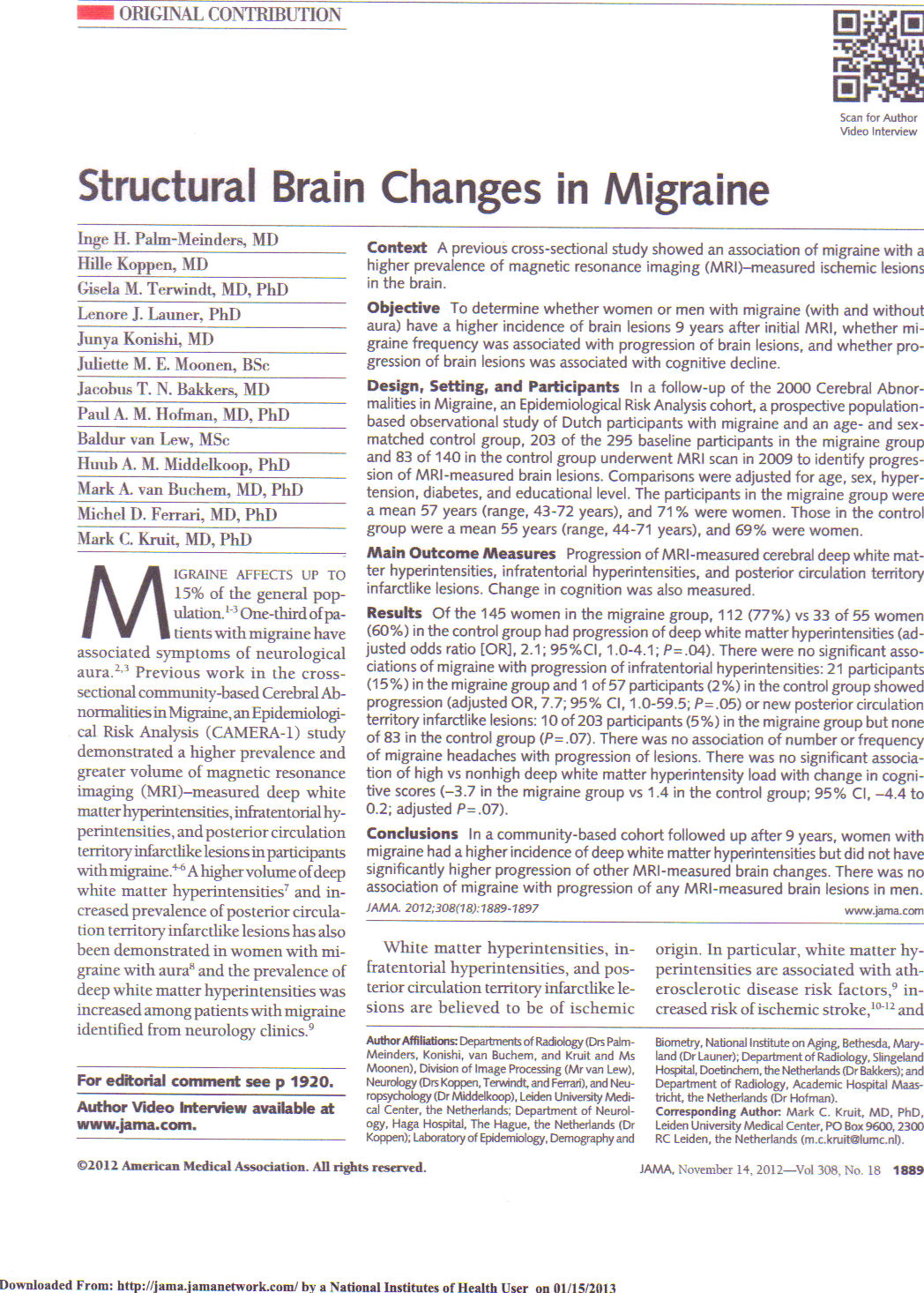
The CAMERA 2 study, the topic for this journal club, follows up these subjects nine years later, looking at progression of their MRI abnormalities and their scores on a battery of cognitive tests performed at the end of the study period.
Journal Review
There were 203 of the 295 original migraineurs and 83 of the original 140 controls available for this second study. Non-participation was equally likely in both groups, and the most commonly cited reasons were lack of interest and difficulty travelling. (The study goes into analysing the non-responders in appropriate detail).
Migraine was diagnosed by standardised International Headache Society criteria. The use of preventatives (that could be protective against vascular changes) or triptans (that theoretically could provoke vascular changes) was probably not prevalent enough to affect the results.
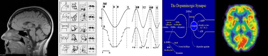
The same imaging protocols were used for the repeat MRI scans, so that there would be a fair comparison over the nine-year period. Analysis of the number and total volume of lesions was done largely by automated software, checked manually by a blinded rater. Abnormalities were grouped into hemispheric T2 weighted deep white matter hyperintensities, infratentorial T2 weighted hyperintensities excluding those hypointense on FLAIR (ie not simply CSF spaces), and other infarct-like lesions in the posterior circulation territory.
Cognitive scores were measured on a number of tests and then converted to Z-scores so that they could be normalised to give an aggregate score for a patient. Association between deep white matter hyperintensity load and follow-up cognitive tests, or between deep white matter hyperintensity load and change in cognition was assessed by linear regression, adjusting for age, sex and educational level. A second linear regression model also adjusted for presence or absence of migraine to see the influence of migraine on the lesion load cognition relationship.
In women only, there was an increased deep white matter hyperintensity load in migraineurs vs controls – 0.02 ml vs 0.00 ml at baseline and 0.09 ml vs. 0.04 ml at follow-up. There was also higher incidence of progression, defined as >0.01 ml volume (77% vs 60%; p=0.02). These were new lesions rather than enlarging pre-existing lesions. Finally there was an increased incidence of “high” progression (23% vs 9%; p=0.03). There was no association with migraine severity measures or its treatment.
There was no effect of presence of migraine on progression of periventricular white matter hyperintensities, or infratentorial hyperintensities or posterior territory infarcts.
The cognitive performances across a number of tests were normalised by calculating Z-scores. For non-statisticians among us, these work like IQ scores, i.e. scores that can be directly compared even if the tests are different. A Z-score of 1.0 means that the patient’s score is one standard deviation better than the average score of the population. This is equivalent to an IQ of about 115 on most tests. (One standard deviation is defined such that around 70% of scores will fall between Z-scores of -1.0 and 1.0.)
Taking all the high-lesion load subjects (defined simply as the worst quintile) vs all the low-lesion load subjects (the remainder), the high lesion load ones had an overall mean Z-score of -3.7 and the low lesion load ones had a mean of 1.4. This was said to be not statistically significant (p=0.07).
(This fooled me at first, possibly not a hard thing to do. A Z-score of -3.7 would mean the high lesion load patients were on average in the bottom sub 1%! But what the authors did was simply add the individual Z-scores across 13 individual tests – they did not take the overall mean across the 13 tests. So the mean for high lesion scores is actually only 0.28 standard deviations lower than the population mean. In fact their “population” is simply all their patients, so if the high lesion load patients were lower than average, the low lesion load patients would have to be higher than average, though by less in magnitude, since there were four times as many low lesion load patients. Statistically, simple addition is fine as one would otherwise just be dividing everything by 13 and that would not change the test results.)
As I have commented in the review for general readers, though, a p-value of 0.07 is nevertheless suggestive that there might be an effect, just that it did not reach significance in this study. The presence or absence of migraine did not influence the lesion-load effect, which had a slightly more reassuring p-value of 0.3, but again if the first effect is really borderline, I am not sure how the linear regression model they use would be expected to behave when adding in the migraine factor.
A general limitation of the study, upon which the authors comment, is that the recording of lesions is rather semi-quantitative and the confidence intervals for odds ratios are wide, suggesting wide inter-subject variability. For example infratentorial progression was considered non significantly associated with migraine because the p value was 0.05. The odds ratio was 7.7 (7 times more likely for progressive lesions if migraine), but the confidence interval was 1.0 to 59.9, meaning that the lower limit was just at the level of no excess risk at all.
Other studies have in fact shown a significant association between lesion load and general cognitive function in apparently healthy elderly subjects (van der Flier et al., 2005). Most previous studies, however, do not show a significant association between migraine itself and declining cognitive function.
The suggestive lesion load effect was not present for lesion load at baseline, only for lesion load at the 9 year follow-up; subjects with a high lesion load 9 years ago did not have a greater change in cognitive function (-0.5 for high load, 0.2 for low lesion load; p=0.4).
In summary, it seems that at a mean age of 57, despite the fact that female migraineurs have scans whose lesions progress more, lesion load per se is associated with (almost significantly) lower cognition, and the presence of migraine does not seem to tighten this possible association. The lesion load 9 years earlier in those of mean age 47 does not predict worse cognition.
Probably this is all indicating that migraine is one of many factors that can result in white matter lesions and some but not all of these factors are associated in turn with cognitive impairment. One factor that is likely to be associated is age; lesions present when subjects were 9 years younger do not predict future impairment, but there is a suggestion that the lesions one has accumulated at a mean age of 57 might be associated with impairment.
In other words, while in general white matter lesions might be associated with impaired cognition, there is no evidence that the white matter lesions seen in younger patients with migraine are going to be associated with impaired cognition in around 10 years time. This is perhaps reflective of the fact that in migraineurs the white matter lesions tend to be small, and to remain small though more numerous over time – perhaps a different natural history from ischaemic lesions that become larger and more confluent as the volume load increases over time.
Conclusions
There is no clear association in this study between migraine and the development of cognitive deficits. There was a significant, but possibly modest, progression in lesion load on MRI compared to the normal aging process. While there was no clear association between lesion load and cognitive deficits, the wide variability in lesion load and the detailed statistical findings indicate that the study is not powered sufficiently to conclude that it is unlikely that lesion load is associated with cognitive deficits. It therefore remains unclear what it is about migraine that results in this excess lesion load but not in cognitive decline, and for us to be completely confident that there is no age range or other subgroup of patients with migraine where such lesions have any clinical significance. As a result, it still remains unclear how we should advise patients with migraine and MRI lesions regarding cerebrovascular preventive measures.

Pingback: Journal Club General Reader Review: Structural Brain Changes in Migraine (the CAMERA-2 Study) | Neurology Online Journal Club